


Cardiac health in cats often goes unnoticed until symptoms become severe, making early detection crucial for effective treatment. Veterinary cardiology has advanced significantly, with cardiac ultrasound emerging as the gold standard for diagnosing heart conditions in felines. This non-invasive imaging technique allows veterinarians to visualise the heart’s structure and function in real-time, providing invaluable information about potential abnormalities. Understanding how this procedure is performed helps pet owners prepare their cats and appreciate the diagnostic value it offers in managing cardiovascular disease.
What is a feline cardiac ultrasound ?
Definition and technical overview
A feline cardiac ultrasound, also known as an echocardiogram, is a diagnostic imaging procedure that uses high-frequency sound waves to create detailed images of a cat’s heart. Unlike X-rays, which provide static images of bone and tissue density, ultrasound technology captures moving images of the heart chambers, valves, and surrounding blood vessels. The procedure is entirely non-invasive and painless, making it ideal for assessing cardiac function without causing distress to the animal.
Types of cardiac ultrasound techniques
Veterinary cardiologists employ several ultrasound modalities during feline cardiac examinations:
- Two-dimensional echocardiography: provides cross-sectional views of cardiac structures
- M-mode echocardiography: offers precise measurements of chamber dimensions and wall thickness
- Doppler echocardiography: evaluates blood flow velocity and direction through heart valves
- Colour Doppler imaging: visualises blood flow patterns using colour-coded representations
These complementary techniques work together to provide a comprehensive assessment of cardiac health, enabling veterinarians to detect conditions ranging from hypertrophic cardiomyopathy to congenital heart defects. The diagnostic power of cardiac ultrasound becomes particularly evident when examining specific heart conditions.
The importance of ultrasound for cardiac diagnosis
Early detection of heart disease
Cardiac ultrasound plays a pivotal role in identifying heart disease before clinical symptoms manifest. Cats are particularly adept at hiding illness, and heart conditions often progress silently until reaching advanced stages. Echocardiography can detect subtle changes in myocardial thickness, chamber size, and valve function that may indicate developing disease. This early detection capability allows for timely intervention, potentially slowing disease progression and improving quality of life.
Common feline cardiac conditions diagnosed
| Condition | Prevalence | Key ultrasound findings |
|---|---|---|
| Hypertrophic cardiomyopathy | Most common (affects 15% of cats) | Thickened left ventricular walls |
| Dilated cardiomyopathy | Less common | Enlarged chambers, reduced contractility |
| Restrictive cardiomyopathy | Moderate prevalence | Impaired diastolic function, atrial enlargement |
| Congenital defects | Rare | Structural abnormalities present from birth |
The diagnostic accuracy of cardiac ultrasound far exceeds that of physical examination or radiography alone, making it indispensable for definitive cardiac diagnosis. Successfully identifying these conditions requires proper preparation of the feline patient.
Preparing the cat for the ultrasound
Pre-examination requirements
Most cardiac ultrasounds require minimal preparation from the owner’s perspective. Cats typically do not need to fast before the procedure, as sedation is rarely necessary for cooperative patients. However, owners should inform the veterinary team about any medications their cat is currently taking, as certain drugs may affect heart function and influence the interpretation of results. If the cat is particularly anxious or aggressive, the veterinarian may recommend a mild sedative to ensure both patient comfort and image quality.
Creating a calm environment
Veterinary practices employ various strategies to minimise stress during cardiac examinations:
- Using feline-friendly handling techniques that respect the cat’s natural behaviours
- Maintaining a quiet examination room with dimmed lighting
- Allowing the cat time to acclimate to the environment
- Permitting owners to remain present when appropriate
- Applying synthetic feline pheromones to reduce anxiety
Some practices offer specialised cat-only appointment times to eliminate the stress of encountering dogs in the waiting area. These preparatory measures help ensure the cat remains sufficiently calm for the actual examination procedure.
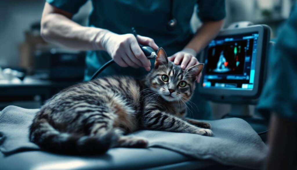
The process of performing a cardiac ultrasound on a cat
Positioning and restraint techniques
The examination typically begins with gentle positioning of the cat in lateral recumbency (lying on its side) on a specialised examination table. Many modern veterinary practices use tables with cut-out sections that allow the ultrasound probe to be positioned from beneath the cat, reducing the need for restraint. Veterinary technicians employ minimal restraint techniques, often simply supporting the cat in a comfortable position rather than forcibly holding it down. The right lateral position is usually preferred initially, providing optimal access to the left side of the chest where the heart is most accessible.
Application of ultrasound gel and probe placement
A small area of fur on the cat’s chest is typically clipped to ensure proper contact between the ultrasound probe and skin. Acoustic coupling gel is then applied to this area, which may be slightly warmed to prevent startling the cat. The veterinarian or veterinary cardiologist places the transducer probe against specific locations on the chest wall, known as acoustic windows, which provide the clearest views of cardiac structures. These standard positions include:
- The right parasternal window: offers views of all four heart chambers
- The left apical window: provides optimal Doppler assessment
- The subcostal window: useful when other views are limited
Duration and patient comfort
A comprehensive cardiac ultrasound typically takes between 20 to 45 minutes, depending on the complexity of the case and the cat’s cooperation level. Throughout the examination, the veterinary team monitors the cat’s stress levels and provides breaks if needed. Most cats tolerate the procedure remarkably well, particularly when handled by experienced personnel. The real-time nature of ultrasound allows the cardiologist to make immediate observations and adjust the examination approach as needed. Once the examination is complete, attention turns to interpreting the wealth of information gathered.
What do the ultrasound results reveal ?
Structural measurements and assessments
The ultrasound examination provides quantitative measurements of various cardiac parameters that are compared against breed-specific and weight-adjusted reference ranges. Key measurements include:
| Parameter | What it measures | Clinical significance |
|---|---|---|
| Left ventricular wall thickness | Myocardial dimensions during diastole | Detects hypertrophy or thinning |
| Left atrial size | Chamber enlargement | Indicates volume overload or pressure |
| Fractional shortening | Contractile function percentage | Assesses systolic performance |
| E/A ratio | Diastolic filling patterns | Evaluates relaxation abnormalities |
Functional and haemodynamic information
Beyond structural measurements, cardiac ultrasound reveals how effectively the heart is functioning. Doppler studies assess blood flow velocities through heart valves, detecting regurgitation or stenosis. The examination can identify blood clots within cardiac chambers, a particular concern in cats with cardiomyopathy who are at risk of thromboembolic disease. Wall motion abnormalities, valve dysfunction, and pericardial effusion are all readily apparent on echocardiographic examination. This comprehensive diagnostic information guides the veterinarian in developing an appropriate management strategy.
Post-examination follow-up: steps and advice
Discussing results with your veterinarian
Following the ultrasound, the veterinary cardiologist will review the findings with the cat’s owner, often using images and videos captured during the examination to illustrate specific abnormalities. Owners should feel empowered to ask questions about the diagnosis, prognosis, and treatment options. Understanding the severity of any detected heart disease helps owners make informed decisions about their cat’s care. Some findings may be incidental and require no immediate treatment, whilst others necessitate prompt therapeutic intervention.
Treatment plans and ongoing monitoring
Depending on the ultrasound findings, the veterinarian may recommend:
- Cardiac medications such as beta-blockers, ACE inhibitors, or diuretics
- Dietary modifications to support cardiovascular health
- Activity restrictions for cats with severe disease
- Regular follow-up examinations to monitor disease progression
- Repeat echocardiography at specified intervals
The frequency of follow-up ultrasounds depends on the specific diagnosis and disease severity, ranging from every few months for rapidly progressing conditions to annually for stable patients. Owners play a crucial role in monitoring their cats at home for signs of deterioration, including increased respiratory rate, reduced activity, or decreased appetite.
Cardiac ultrasound represents an essential diagnostic tool in modern feline medicine, offering unparalleled insight into heart structure and function. Through careful preparation, skilled execution, and thorough interpretation, this non-invasive procedure enables early detection and effective management of cardiovascular disease in cats. Regular cardiac screening, particularly for breeds predisposed to heart disease or cats with cardiac murmurs, can significantly improve outcomes and quality of life for our feline companions.